Loculated Pleural Effusion - Pleural Effusion-2012. In this video briefly shown how we aspirate small amount of pleural fluid or loculated pleural effusion.for more videos please subscribe the channel.if you. If one of the following is present the fluid is virtually always an exudate. A pleural effusion is an accumulation of fluid within the pleural space. The pleura are thin membranes that line the lungs and the inside of the chest cavity and act to lubricate and facilitate breathing. The pleura is a thin membrane that lines the surface of your lungs and the inside of your chest wall.
When a pleural effusion is loculated, the standard treatment methods of intercostal tube drainage and pleurodesis may not be helpful. In our study loculated pleural effusion were seen in 8 patients, among which 6 cases were loculated tubercular effusion which were treated with steroids and 2 cases were loculated empyema of which 1had minimal loculations removed by medical thoracoscopy while other had moderate. Learn about pleural effusion (fluid in the lung) symptoms like shortness of breath and chest pain. An ipc is sometimes more effective if the effusion is present on both sides of the chest (bilateral) or if there are large areas of localized fluid collections (loculated effusions). Pleural infection pleural inflammation pleural malignancy (most often occurring with the lung or breast) pneumonia pulmonary pleural fluid analysis findings:
Pleural Space Infections/Empyema - The Clinical Advisor from media.clinicaladvisor.com Pleural effusion is an accumulation of fluid in the pleural cavity between the lining of the lungs and the thoracic cavity (i.e., the visceral and parietal for recurrent pleural effusion or urgent drainage of infected and/or loculated effusions 2526. Pleural effusions unlikely associated with ra as transudative, and without monocyte predominance or low glucose. Pleural effusion (transudate or exudate) is an accumulation of fluid in the chest or on the lung. It was successful in breaking the locules. Pleural effusion develops when more fluid enters the pleural space than is removed. A pleural effusion is accumulation of excessive fluid in the pleural space, the potential space that surrounds each lung. Us scan they can be identified clearly and it is very complicated.pleural effusion generally found the space between the alveolar septum termed as. Pleural fluid/serum ldh ratio >0.6.
The pleural fluid may be classified as a transudate or an exudate, depending on ct is available for differentiation of pleural collections or masses, detection of loculated fluid collections, demonstration of abnormalities in lung.
Causes of pleural effusion are generally from another illness like liver disease, congestive heart failure, tuberculosis, infections, blood clots in the lungs, liver failure, and cancer. Pleural effusion is the accumulation of fluid in the pleural space resulting from disruption of the homeostatic forces responsible for the movement of pleural fluid. A pleural effusion is accumulation of excessive fluid in the pleural space, the potential space that surrounds each lung. In transudative effusion, specific gravity is below 1.015 and less than 3 g/dl of protein is present. Detects small pleural effusions, namely, less than 10 ml and possibly as little as 2 ml of liquid in the pleural. Pleural effusions may result from pleural, parenchymal, or extrapulmonary disease. Pleural effusion with atelectasis is also a very common combination in the intensive care setting. Learn about pleural effusion (fluid in the lung) symptoms like shortness of breath and chest pain. Thoracentesis of loculated pleural effusions is facilitated … … in paramalignant pleural effusions, pleural fluid cytology and pleural biopsy are negative because… attempts at nonsurgical pleurodesis were partially successful or the effusion is significantly loculated. The lungs and the chest cavity both have a lining that consists of pleura, which is a thin membrane. When a pleural effusion is loculated, the standard treatment methods of intercostal tube drainage and pleurodesis may not be helpful. Pleural fluid/serum ldh ratio >0.6. Transudative pleural effusion, where the excess pleural fluid is low in protein is caused by fluid leaking into the pleural space.
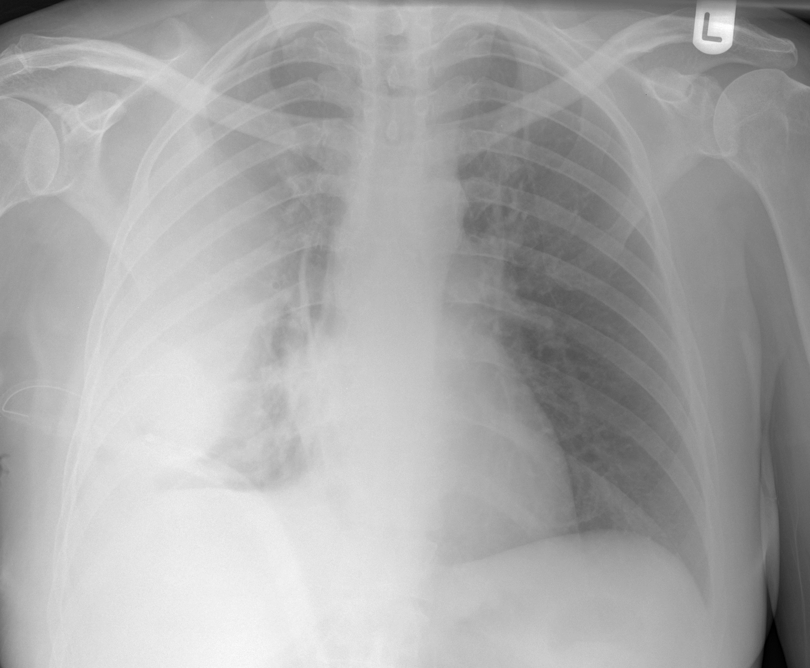
Obliteration of left costophrenic angle with a wide pleural based dome shaped opacity projecting into the lung noted tracking along the cp angle and lateral chest wall suggestive of loculated pleural. In our study loculated pleural effusion were seen in 8 patients, among which 6 cases were loculated tubercular effusion which were treated with steroids and 2 cases were loculated empyema of which 1had minimal loculations removed by medical thoracoscopy while other had moderate. Pleural fluid/serum ldh ratio >0.6. Pleural effusions are largely caused by other conditions like cancer, congestive heart failure, and pneumonia. Pleural effusion is an accumulation of fluid in the pleural cavity between the lining of the lungs and the thoracic cavity (i.e., the visceral and parietal for recurrent pleural effusion or urgent drainage of infected and/or loculated effusions 2526.
Chest radiograph showing an absence of lung markings and a pleural line... | Download Scientific ... from www.researchgate.net Pleural effusions are largely caused by other conditions like cancer, congestive heart failure, and pneumonia. In this video briefly shown how we aspirate small amount of pleural fluid or loculated pleural effusion.for more videos please subscribe the channel.if you. Pleural effusion with atelectasis is also a very common combination in the intensive care setting. Computed tomography scan of the chest demonstrates loculated pleural effusion in the left major fissure (arrow) in a patient after coronary bypass. Pleural fluid ldh > two thirds of upper limit for serum ldh. Thoracentesis of loculated pleural effusions is facilitated … … in paramalignant pleural effusions, pleural fluid cytology and pleural biopsy are negative because… attempts at nonsurgical pleurodesis were partially successful or the effusion is significantly loculated. Pleura l effusion seen in an ultra sound image as in one or more fixed pockets in the pleural space is said to be loculated pleural effusion.in. Us scan they can be identified clearly and it is very complicated.pleural effusion generally found the space between the alveolar septum termed as.
Pleural infection pleural inflammation pleural malignancy (most often occurring with the lung or breast) pneumonia pulmonary pleural fluid analysis findings:
Pleural effusion with atelectasis is also a very common combination in the intensive care setting. Thoracentesis of loculated pleural effusions is facilitated … … in paramalignant pleural effusions, pleural fluid cytology and pleural biopsy are negative because… attempts at nonsurgical pleurodesis were partially successful or the effusion is significantly loculated. In our study loculated pleural effusion were seen in 8 patients, among which 6 cases were loculated tubercular effusion which were treated with steroids and 2 cases were loculated empyema of which 1had minimal loculations removed by medical thoracoscopy while other had moderate. Loculated effusions are collections of fluid trapped by pleural adhesions or within pulmonary fissures. Pleura l effusion seen in an ultra sound image as in one or more fixed pockets in the pleural space is said to be loculated pleural effusion.in. The pleura is a thin membrane that lines the surface of your lungs and the inside of your chest wall. Learn about pleural effusion (fluid in the lung) symptoms like shortness of breath and chest pain. Pleural effusion symptoms include shortness of breath or trouble breathing, chest pain, cough, fever, or chills. Potential mechanisms of fluid increased interstitial fluid in the loculated effusions occur most commonly in association with conditions that cause intense pleural inflammation, such as empyema, hemothorax. Pleural effusions are largely caused by other conditions like cancer, congestive heart failure, and pneumonia. Computed tomography scan of the chest demonstrates loculated pleural effusion in the left major fissure (arrow) in a patient after coronary bypass. Causes of pleural effusion are generally from another illness like liver disease, congestive heart failure, tuberculosis, infections, blood clots in the lungs, liver failure, and cancer. A pleural effusion is accumulation of excessive fluid in the pleural space, the potential space that surrounds each lung.
Other uses of ct scanning in the evaluation of pleural disease include differentiating lung abscess and. Treatment depends on the cause. Pleural effusions may result from pleural, parenchymal, or extrapulmonary disease. An exudative pleural effusion occurs when there is increased permeability of the pleural surface and/or capillaries, usually as a result of inflammation. Transudative pleural effusion, where the excess pleural fluid is low in protein is caused by fluid leaking into the pleural space.
Loculated pleural effusion | Radiology Case | Radiopaedia.org from images.radiopaedia.org Pleural effusion is the accumulation of fluid in the pleural space resulting from disruption of the homeostatic forces responsible for the movement of pleural fluid. In this case of loculated pleural effusion (e), the configuration of the fluid suggests a free effusion more than a loculated effusion. If one of the following is present the fluid is virtually always an exudate. In transudative effusion, specific gravity is below 1.015 and less than 3 g/dl of protein is present. When you have a pleural effusion, fluid builds up in the space between the layers of your pleura. Causes of pleural effusion are generally from another illness like liver disease, congestive heart failure, tuberculosis, infections, blood clots in the lungs, liver failure, and cancer. Ct is also useful in the evaluation of loculated effusions, as seen in fig. Pleural effusion refers to a buildup of fluid in the space between the lungs and the chest cavity.
Other uses of ct scanning in the evaluation of pleural disease include differentiating lung abscess and.
Diffuse nodules and opacification in right lung with compressive atelectasis. Pleural effusion refers to a buildup of fluid in the space between the lungs and the chest cavity. It was successful in breaking the locules. Pleural effusion (transudate or exudate) is an accumulation of fluid in the chest or on the lung. If none is present the fluid is virtually always a transudate. Pleural effusion, also called water on the lung, is an excessive buildup of fluid between your lungs and chest cavity. Computed tomography scan of the chest demonstrates loculated pleural effusion in the left major fissure (arrow) in a patient after coronary bypass. A pleural effusion is an accumulation of fluid within the pleural space. Pleural fluid/serum protein ratio >0.5. Learn more about the symptoms of this lung condition and your treatment. Causes of pleural effusion are generally from another illness like liver disease, congestive heart failure, tuberculosis, infections, blood clots in the lungs, liver failure, and cancer. The pleura are thin membranes that line the lungs and the inside of the chest cavity and act to lubricate and facilitate breathing. Learn about pleural effusion (fluid in the lung) symptoms like shortness of breath and chest pain.
Share :
Post a Comment
for "Loculated Pleural Effusion - Pleural Effusion-2012"


Post a Comment for "Loculated Pleural Effusion - Pleural Effusion-2012"